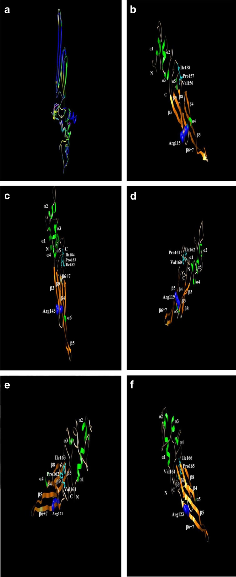
Fig. 2.
Structure analyses of five small heat shock proteins from C. suppressalis. a Homology modeling analyses of the CsHSP19.8 (yellow), CsHSP21.4 (blue), CsHSP21.5 (cyan), CsHSP21.7a (purple), and CsHSP21.7b (green) with human αB-crystallin V* structure (PDB ID: 2YGD) (white) as template. b–e The α-helix shown in yellow and β-strand shown in orange. The conserved Arg is shown in blue sphere and the V/IXI/V motif is shown in cyan stick and ball. b CsHSP19.8, c CsHSP21.4, d CsHSP21.5, e CsHSP21.7a, and f CsHSP21.7b

