Figure 1.

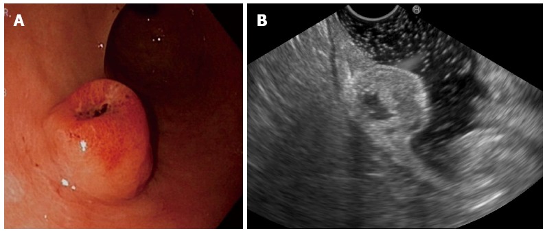
Endoscopic view. A: Showing a subepithelial lesion with ulceration and blood clotting; B: Showing a hypoechoic lesion arising from the second and third layers of the gastric wall, with a central anechoic area.

Endoscopic view. A: Showing a subepithelial lesion with ulceration and blood clotting; B: Showing a hypoechoic lesion arising from the second and third layers of the gastric wall, with a central anechoic area.