Figure 1.
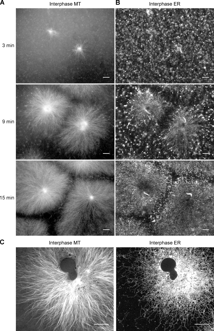
ER network formation in crude interphase X. laevis egg extracts. Demembranated sperm was added to a crude interphase X. laevis egg extract containing Alexa fluor 488–labeled tubulin and the hydrophobic dye DiIC18. The formation of MT asters (A) and ER network (B) was followed over time by confocal fluorescence microscopy (for full time course, see Videos 1 and 2). Sperm was added at time zero. Bars, 30 µm. (C) As in A and B, but after 60 min, a time point at which the NE has formed. Bars, 20 µm.

