Figure 7.
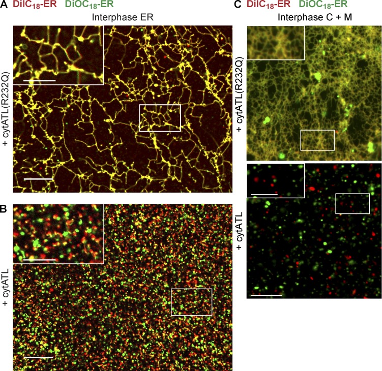
ATL mediates the fusion of membranes into an ER network. (A) A crude interphase extract was preincubated for 5 min with 2 µM of an inactive mutant of the cytosolic fragment of ATL (ATL(R232Q)). Then, membranes were added that were separately prestained with DiIC18 (red) and DiOC18 (green). The sample was imaged after 30-min incubation by confocal microscopy with a short exposure time (40 ms). (B) As in A, but with wild-type cytosolic fragment of ATL (cytATL). Insets in A and B show a magnified view of the boxed areas. (C) As in A and B, but with membrane-depleted interphase cytosol and a mixture of light membranes that were prestained with either DiIC18 or DiOC18. Insets show a magnified view of the boxed areas. Bars: (A and B) 20 µm; (A and B, insets) 10 µm; (C) 10 µm; (C, insets) 5 µm.

