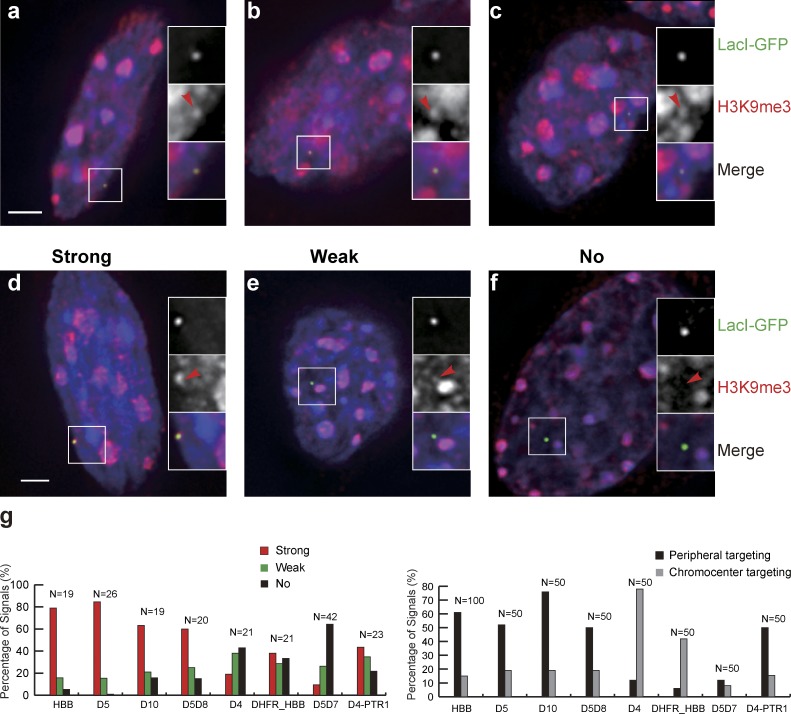
Figure 5.
Differential targeting to the nuclear periphery versus chromocenter correlates with H3K9me3 levels over BAC transgenes. (a–f) BAC transgene locations (EGFP-LacI staining), H3K9me3 immunostaining, DAPI (blue). Enlarged insets show regions of magnification with red arrowheads in red channel (middle) pointing to location of transgenes in green (top) channel. (a–c) HBB BAC transgene locations in clone HBB-C3 overlap with H3K9me3 immunostaining foci regardless of whether transgenes are located at nuclear periphery (a), chromocenter (b), or interior (c). (d–f) Examples from clone HBB C3 showing strong (d), weak (e), or no (f) H3K9me3 signals over the BAC transgenes. (g) Strength of H3K9me3 immunostaining over different BAC transgenes (left) correlates with differential targeting of BAC transgenes (right) to nuclear periphery versus chromocenter. Bars, 2 µm. Data shown for each BAC are pooled from at least two independent experiments.

