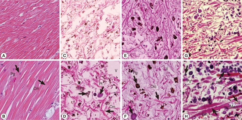
Fig. 1.
The representative tissue of control snails (A, B), snails exposed to camellia (C, D), snails exposed to mangosteen (E, F), and snails exposed to niclosamide (G, H). Hyperpigmentation of pigment cells, increasing numbers of lipid vacuoles, and atrophy of columnar muscle fibers were observed in camellia, mangosteen, and niclosamide-treated groups. Cm, columnar muscle fibers; Pt, protein cells; Pg, pigment cells; V, lipid vacuoles.

