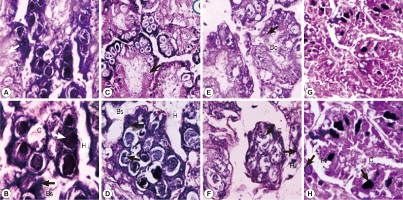
Fig. 3.
Representative digestive glands of control snails (A, B), those exposed to camellia (C, D), those exposed to mangosteen (E, F), and those exposed to niclosamide (G, H). Camellia, mangosteen, and niclosamide-treated groups showed a small number of calcium cells, dilation of the digestive gland, and large hemolymphatic spaces compared with the control snails. Dc, digestive cells; C, calciferous cells; Bs, basophilic cells; H, hemolymphatic spaces between the tubules; Pt, protein cells.

