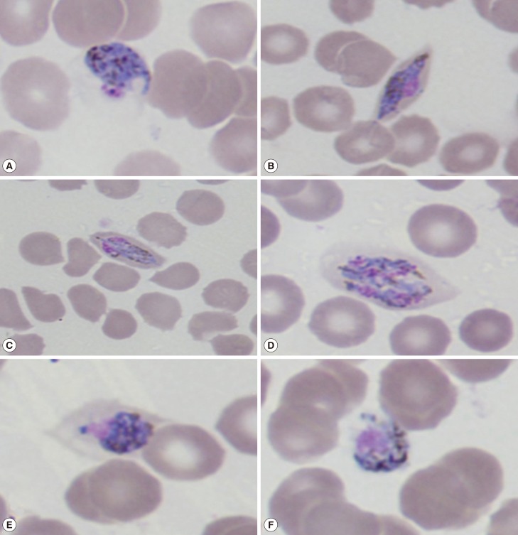
Fig. 1.
Giemsa-stained thin blood smears at the time of presentation (×1,000) showing different life-cycle stages of Plasmodium ovale wallikeri. (A, B) Different types of trophozoites seen in oval RBCs. (C, D) Mature and immature P. ovale schizonts with 4-6 merozoites containing large nuclei seen clustered around a mass of dark-brown pigment. (E, F) Gender-specific round to oval P. ovale gametocytes almost filling the RBCs.

