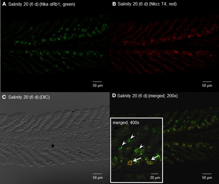
Figure 12.
Immunofluorescent localization of Na+/K+-ATPase α-subunit (Nkaα) and Na+:K+:2Cl− cotransporter (Nkcc) in the gills of Himantura signifer exposed to brackish water (BW; salinity 20) for 6 d after a progressive increase in salinity. Immunofluorescence using (A) anti-NKA αRb1 antibody (green) or (B) anti-NKCC T4 antibody (red). The differential interference contrast image (DIC) is shown in (C). All channels (green and red) are merged and overlaid with DIC in (D), whereby integration of red and green channels resulted in a yellow-orange coloration. In the inset of (D), arrows indicate the staining of Nka and Nkcc in a type of BW ionocyte, while arrowheads denote the staining of only Nka in another type of ionocyte. Magnification: 200× for (A–D), or 400× for inset of (D). Reproducible results were obtained from three individuals.

