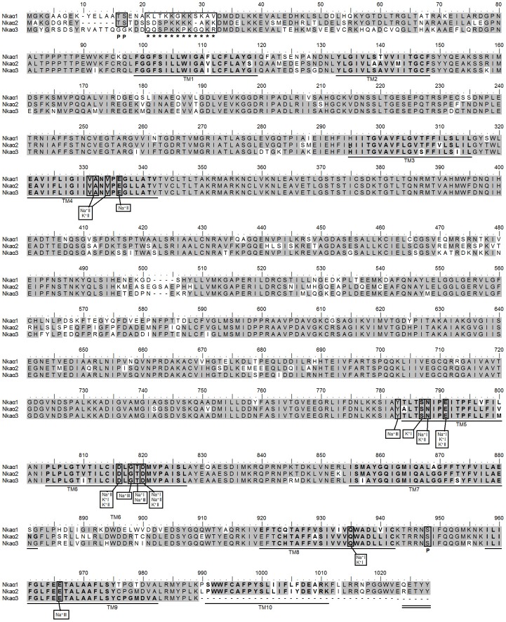
Figure 4.
Molecular characterization of Na+/K+-ATPase (Nka) α1, Nkaα2, and Nkaα3 from the gills of Himantura signifer. A multiple amino acid sequence alignment of Nkaα1, Nkaα2, and Nkaα3 from the gills of H. signifer. Identical amino acid residues are indicated by shaded residues. The ten predicted transmembrane regions (TM1-TM10) are underlined and in bold. Vertical boxes represent coordinating residues for Na+ or K+ binding. “P” denotes phosphorylation sites and asterisks indicate the lysine-rich region. The conserved region containing the KETYY sequence motif is double underlined.

