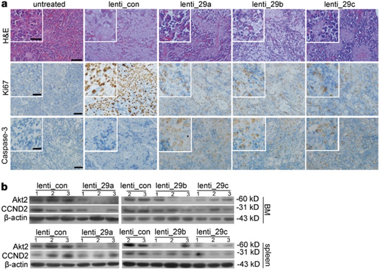
Figure 8.
Histological and immunohistochemical analysis of spleens collected from the experimental NOD/SCID mice and western blot assays of the target proteins. (a) Spleen sections obtained from NOD/SCID mice with different treatments were analyzed for the degree of engraftment by H&E (upper), Scare bar: 50 μm (inset: 20 μm); cell proliferation by Ki-67 staining (middle) and apoptosis by caspase-3 staining (lower), Scare bar: 20 μm (inset: 10 μm). The results of one mouse from each group were shown. (b) Western blotting analysis of Akt2 and CCND2 protein levels in murine BM and spleen. The results of three mice from each group were shown

