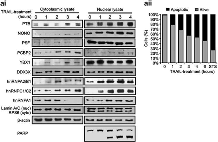
Figure 2.
Relocalization of members of the PTB complex during apoptosis. (ai) Western blotting of nuclear and cytoplasmic fractionated lysates of MCF7 cells treated with TRAIL over a 4 h time course using antibodies against indicated proteins. RPS6 and Lamin A/C antibodies were used as cytoplasmic and nuclear markers, respectively. PARP cleavage was used to indicate apoptosis and β-actin was used as a loading control. (aii) Control and TRAIL-treated MCF7 cells were stained with Annexin V-FITC and propidium iodide at the indicated time points and subjected to FACS analysis. Staurosporin-treated MCF7 cells (STS) served as a positive control at 6 h

