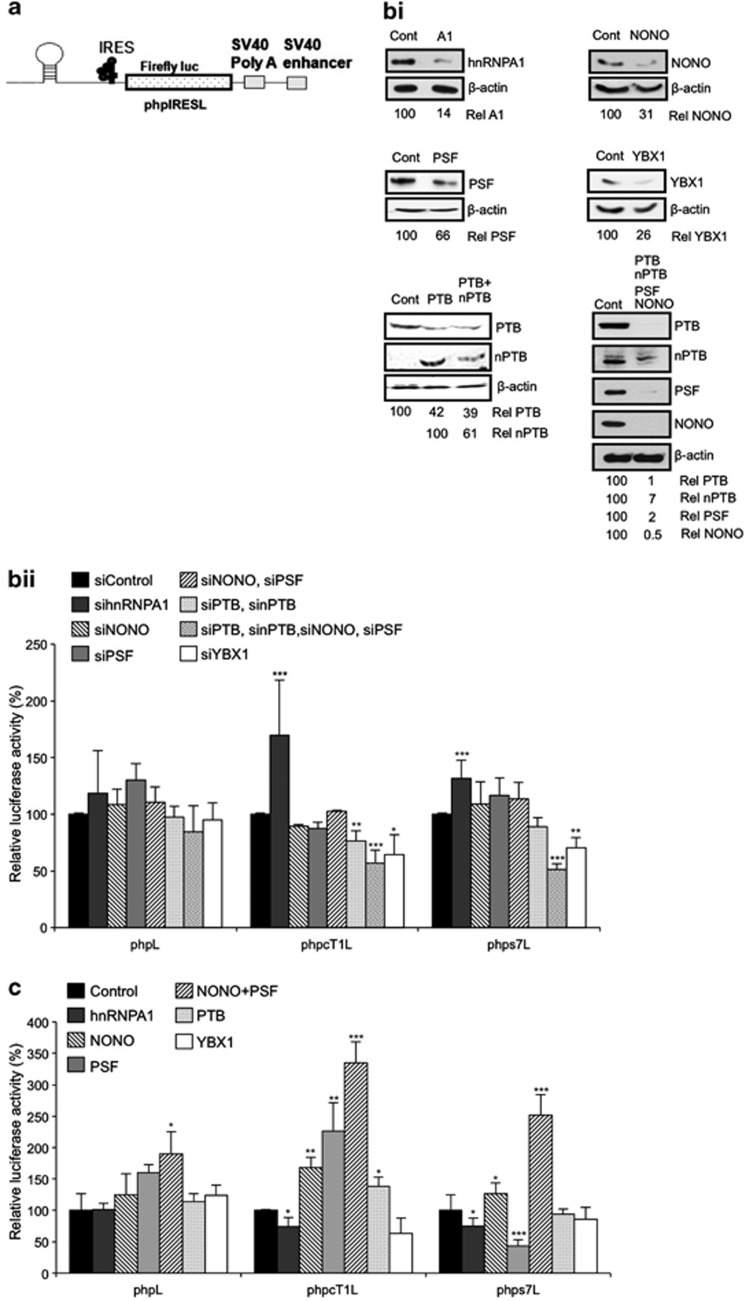
Figure 5.
Altering the levels of PTB-interacting proteins effects IRES activity. (a) Schematic representation of the monocistronic constructs used in figures bii and c, which contains a stable hairpin loop downstream of the 5′ m7G cap in order to inhibit cap-dependent translation of the Firefly luciferase reporter. (bi) HeLa cells were co-transfected with siRNA against the indicated proteins together with the monocistronic reporter plasmid, incubated for 48 h then harvested and western blot analysis was carried out to confirm RNAi success. Immunoblots are quantified relative to β-actin levels, and expressed as a percent of levels in the control siRNA treated lysate. (bii) Luciferase assays were carried out on the RNAi lysates. Data are shown relative to a control experiment using control siRNA. Significance (*P<0.05, **P< 0.01 or ***P<0.005) was calculated using an unpaired two-tailed Student's t-test (n=3), error bars represent S.D. (c) Reticulocyte lysates were primed with 100 ng recombinant protein and 100 ng of in vitro transcribed m7G capped and polyadenylated reporter RNA, incubated at 30 °C for 90 min, then assayed for luciferase activity. Data are shown relative to a control experiment with no recombinant protein added. Significance (*P<0.05, **P<0.01 or ***P<0.005) was calculated using an unpaired two-tailed Student's t-test (n=3), error bars represent S.D.

