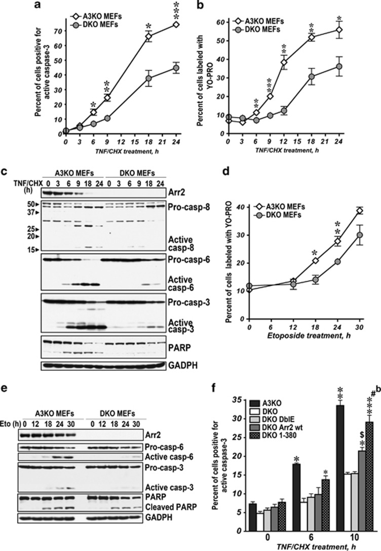
Figure 5.
The generation of 1-380 facilitates apoptotic cell death. (a–c) A3KO and DKO MEFs were treated with TNFα/CHX (10 ng/ml and 10 μg/ml) for the indicated times. Apoptosis was assessed by the percentage of cells positive for active caspase-3 (a) or stained with YO-PRO-1 (b) using FACS in four independent experiments. The data were analyzed by two-way ANOVA with MEF and Time as main factors. In both caspases-3 and YO-PRO experiments, the effects of MEF were highly significant (P<0.0001). ***P<0.001; **P<0.01; *P<0.05 to DKO MEFs according to unpaired Student's t-test for each time point. (c) The activation of caspases in A3KO and DKO MEFs treated with TNFα/CHX analyzed by western blotting. (d) A3KO and DKO MEFs were treated with etoposide (100 μM) for the indicated times. Apoptosis was assessed by the percentage of cells stained with YO-PRO-1. The two-way ANOVA analysis with MEF and Time as main factors yielded highly significant effect of MEF (P=0.0001). **P<0.001; *P<0.005 to DKO MEFs according to unpaired Student's t-test for each time point. (e) The activation of caspase-6 and -3 in A3KO and DKO MEFs treated with etoposide analyzed by western blotting. (f) A3KO or DKO MEFs expressing GFP, wt Arr2+GFP, DblE+GFP, or 1-380+GFP were exposed to TNFα/CHX for the indicated times. Apoptosis was determined as the percentage of cells positive for active caspase-3 in GFP-positive subsets measured by FACS (means±S.E.M. of four experiments). The data were analyzed by one-way ANOVA for each time point with MEF Type as the main factor. The MEF effect was highly significant for 6 and 10 h TNFα/CHX treatment time points. **P<0.001, *P<0.01 to DKO, DKO DblE, and DKO Arr2 wt; ***P<0.001; *P<0.05 to DKO MEFs; $P<0.05, #P<0.001 to DblE-GFP; bP<0.01 to Arr2-GFP according to Bonferroni/Dunn post hoc test. See also Supplementary Figure S3

