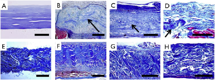
Figure 9.
Extracellular matrix staining (Masson's Trichrome) of unimplanted and implanted samples. Multilayer collagen (A) and Permacol™ (E) patches prior to implant show uniform thickness and distinct morphology. For multilayer collagen implants, wavy morphology collagen (arrows) is noted above muscle (red) and adjoining highly cellularized peritoneal layer at 1 month (B), is clearly delineated in the center of the receullularized implant at 2 months (C), and is seen in isolated pockets at 3 months (D). Permacol™ implants can be clearly distinguished from host tissue at 1 month (F), 2 months (G) and 3 months (H), resembling preimplant structure and morphology. Scale bar A-D: 200 μm and E-H: 500 μm.

