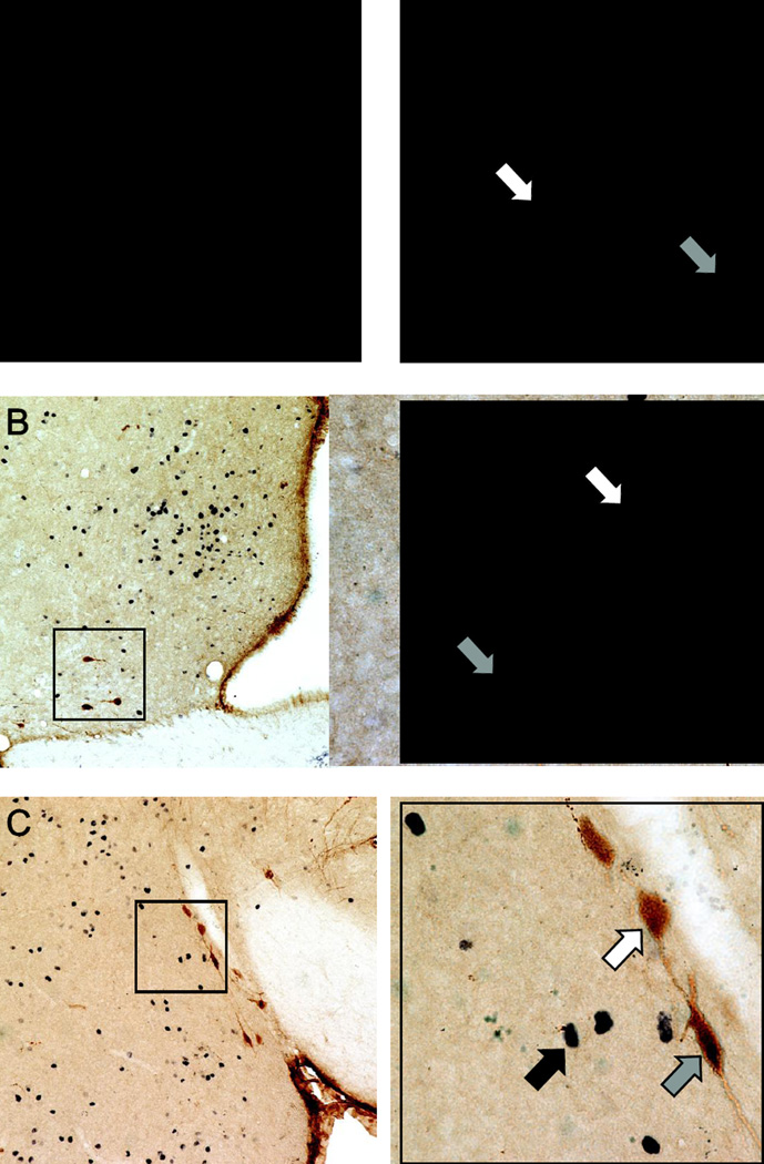
Figure 3.
Immunohistochemical staining for oxytocin and Fos. Representative photomicrographs of double-label immunohistochemistry in sections depicting (A) the paraventricular nucleus of the hypothalamus (PVH), (B) the medial preoptic area (MPOA), and (C) the anterior division of the medial amygdala (MeA). A higher magnification (40X objective) of the areas highlighted within the black boxes is provided on the right of each lower magnification (10X objective) image. White arrows indicate oxytocin-immunoreactive cells (brown cytoplasmic staining). Black arrows indicate Fos-immunoreactive cells (black nuclear staining). Gray arrows indicate cells immunoreactive for both oxytocin and Fos (brown cytoplasmic staining with black nuclear staining). Scale bars = 100 µm (lower magnification) and 25 µm (higher magnification).

