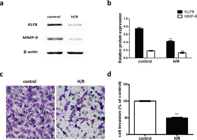
Figure 6.
KLF8 and MMP-9 protein levels and trophoblast invasion in H/R-exposed HTR-8/SVneo cells. A, Representative Western blot for KLF8 and MMP-9 protein expression in HTR-8/SVneo cells exposed to H/R. β-Actin was used as an internal control for protein loading. B, The relative amounts of KLF8 and MMP-9 proteins were standardized by β-actin levels. The data are expressed as the mean ± standard error of the mean from 3 separate experiments. **P < .01 (compared with control). C, Representative image shows HTR-8/SVneo cells passing through the membrane. Control, nontreated cells were maintained under normal conditions for 48 hours. B, H/R cells were cultured under hypoxic conditions (2% oxygen) for 8 hours, followed by reoxygenation for 16 hours, and the H/R intervention described above was repeated twice. D, The histogram represents the relative numbers of migrating cells (%). **P < .01 (compared with the control group). H/R, hypoxia–reoxygenation; KLF8, Krüppel-like factor 8; MMP-9, metalloproteinase 9.

