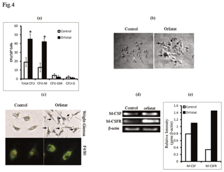
Figure 4. Orlistat augments differentiation of BMDM associated with increased expression of M-CSF and M-CSFR.
BMC (1x10⁴ cells/ml) harvested from control or orlistat-administered tumor-bearing mice were cultured in vitro in the presence of L929 culture medium (20%v/v) as a source of M-CSF, for 10 days to allow the BMC to differentiate into colonies. Colonies were counted based on cellular morphology of each colony forming unit (CFU) displaying features of CFU-M, CFU-GM and CFU-G phenotype (a). CFU-M obtained from the BMC of control group displayed lesser number of Mϕ-like cells compared to orlistat-treated group where the colonies were denser with larger macrophage like cells, as indicated by arrows (b). BMC (1x10⁶ cells/ml) obtained from control or orlistat-administered tumor-bearing mice were also processed for RT-PCR to detect expression of mRNA for MCSF and M-CSFR. Bars shown in (e) are densitometric scan of bands shown in (d), which are from a representative experiments out of 3 experiments with similar results. BMDM grown on glass cover slips in petri-dishes were stained with Wright Giemsa stain (c upper panel) and F4/80 FITC-conjugated antibody (c lower panel). As indicated by arrows BMDM of orlistat administered group showed increased size, spreading and cytoplasmic extensions. Plates shown are from a representative experiment. Values shown in (a) are mean ± SD of three independent experiments done in triplicate.*p<0.05 vs. values of respective control.

