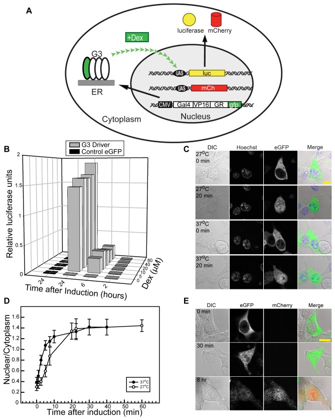
Figure 1. G3U inducible system in mammalian cell culture.
(A) Diagram of G3U system using luciferase (pCMV:G3 and pUAS:Luciferase) or mCherry reporters (top, pCMV:G3 and pUAS:mCherry). A CMV promoter drives transcription of a chimeric transcription factor, G3, which encodes contains a GAL4 DNA binding domain, the VP16 transcription activation domain, a rat glucocorticoid receptor binding domain (GR) and eGFP. Synthesized G3 protein localizes to the cytosol. Dex treatment triggers the dimerization of G3, which translocates into the nucleus. Nuclear G3 activates a second construct containing five tandem repeats of the UAS sequence upstream of the hsp70 minimal promoter. The reporter gene (luciferase or mCherry) is under the control of this system. (B) Luciferase assay of G3U inducible system in cell culture. HEK293T cells transfected with pCMV:G3 and pUAS:Luciferase were lysed and luciferase activity measured at different concentrations (0-80 µM) and treatment durations (2-24 hr) of Dex. Relative luciferase activity is plotted as a function of duration and Dex concentration. (C) Live cell imaging of G3 translocation after induction. HEK293 cells were transfected with pCMV:G3 and pUAS:mCherry and induced with 10 μM Dex at 27°C and 37°C. Confocal images were taken before and 20 min after induction; G3 (eGFP), nucleus (Hoechst). Scale bar is 10 μm. (D) Nuclear translocation rate of G3 in HEK293T cells at different temperatures. Fluorescence intensity was measure in nuclear and cytoplasm of live 293T cells at 27°C and 37 °C. Dex (10 μM) was added and mixed into medium. Error bars represent standard deviation (n = 17 at 37 °C and n = 15 at 27 °C). (E) mCherry reporter expression after induction. HEK293T cells transfected with pCMV:G3-GFP and pUAS:mCherry were induced with 10 μM Dex and fixed at different times after induction. G3 (eGFP) and mCherry images show the movement of G3 and expression of mCherry. Scale bar is 10 μm.

