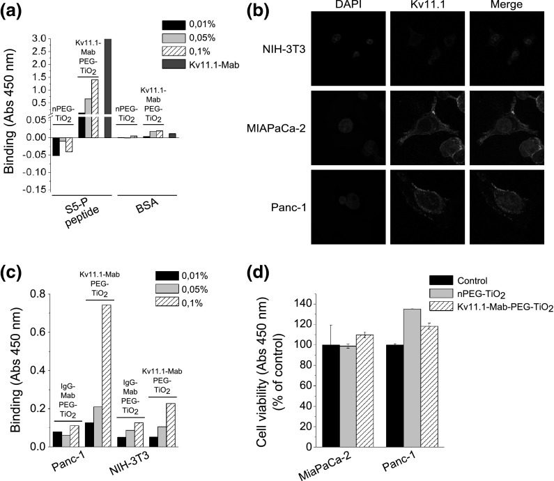
Fig. 4.
NPs’ interaction with PDAC cells: a binding specificity of Kv 11.1-Mab-PEG–TiO2 NPs determined through an ELISA assay performed on the S5P peptide; b Kv 11.1 expression in NIH-3T3, MIAPaCa-2, and Panc-1 cells. Immunofluorescence was performed on cells seeded onto glass slides coated with 20 μg/mL Fibronectin, by means of Kv 11.1-Mab and Alexa-488-conjugated secondary antibody. Photographs were taken using a C1 confocal microscope (Nikon); c binding of Kv 11.1-Mab-PEG–TiO2 NPs on cells: MIAPaCa-2 and NIH-3T3 cells were treated with different concentrations of Kv 11.1-Mab-PEG–TiO2 and IgG-Mab-PEG–TiO2; d cell viability (WST-1) assay performed on MIAPaCa-2 and Panc-1 cells in the presence of either PEG–TiO2 or Kv 11.1-Mab-PEG–TiO2 NPs, both at 0.1 % concentration

