
Figure 4.
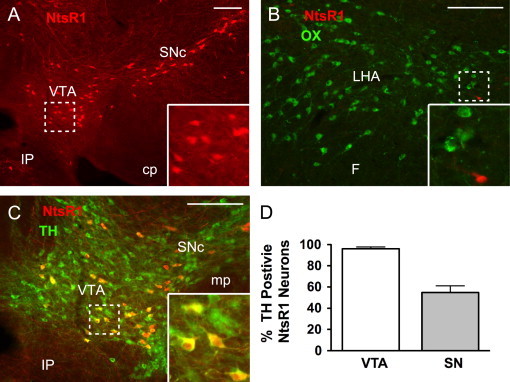
Visualization of NtsR1 neurons in the brain. Transgenic mice expressing cre recombinase in NtsR1 neurons (NtsR1cre mice) were crossed to ROSA26-tdTomato reporter mice to express Tomato in NtsR1 neurons (red). (A) NtsR1 neurons are found within the midbrain, including the VTA and SNc. (B) Few NtsR1 neurons are found in the LHA, none of which co-localize with OX neurons (green). (C) Many of the NtsR1 neurons in the midbrain colocalize with TH (green), a marker of DA neurons. (D) Average percentage of NtsR1 neurons in the VTA and SN that co-express TH. Error bars depict±SEM. Scale bars=100 µm. Dashed boxes identify regions that are digitally enlarged within each panel. VTA=ventral tegmental area, SNc=substantia nigra compacta, IP=interpeduncular nucleus, cp=cerebral peduncle, LHA=lateral hypothalamic area, F=fornix, mp=mammillary peduncle. (For interpretation of the references to color in this figure legend, the reader is referred to the web version of this article.)