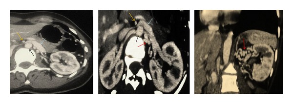
Figure 2.

CTA of the abdomen and pelvis showed complete disappearance of the renal vein thrombosis and demonstrated the nutcracker syndrome: compression of the LRV between the aorta (black arrow) and the SMA (yellow arrow), with a dilatation of the hilar portion of the LRV (blue arrow). The red arrow showed and surrounding vascular collaterals.
