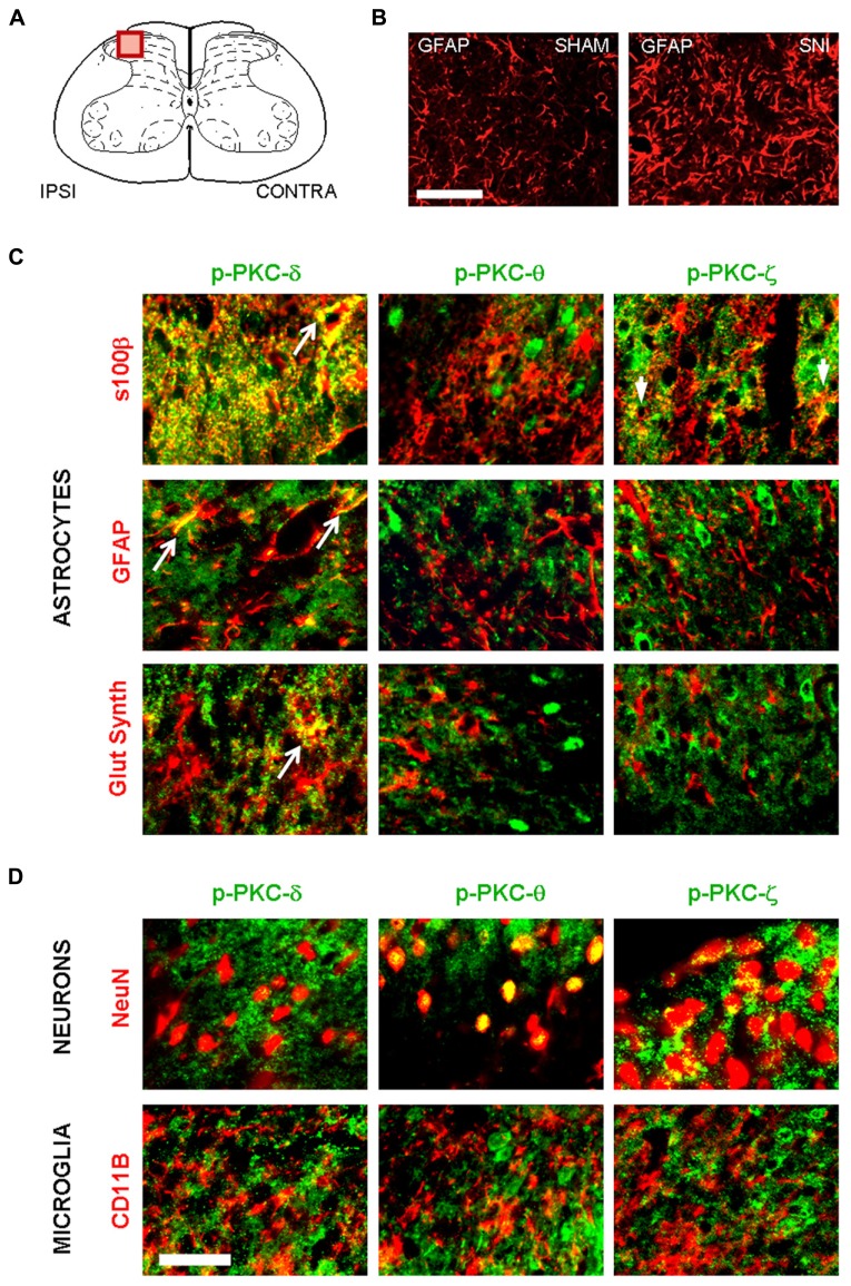
FIGURE 6.
Following peripheral nerve injury, astrocytes are the source of PKC-δ activation. (A) Schematic showing the region of interest in the spinal dorsal horn. (B) Immunofluorescence highlighting spinal GFAP immunoreactivity one week after sham (left picture) or in SNI surgery (right picture). Scale bar 100 μm. (B,C) Immunofluorescence detecting isoforms of phospho PKC. (C) A colocalization is found between markers for astrocytes (s100β, GFAP and glutamine synthetase, red) and activated PKC-δ and -ϖ (green, first and third columns). No overlap was found between phospho PKC θ and astrocytes. (D) Phospho-PKC-θ and -ϖ, but not phospho-PKC-δ colocalizewith neuronal marker (NeuN, upper images). No isoform could be detected in microglia (CD11b, lower images). Scale bar, 100 μm.

