
Figure 4.
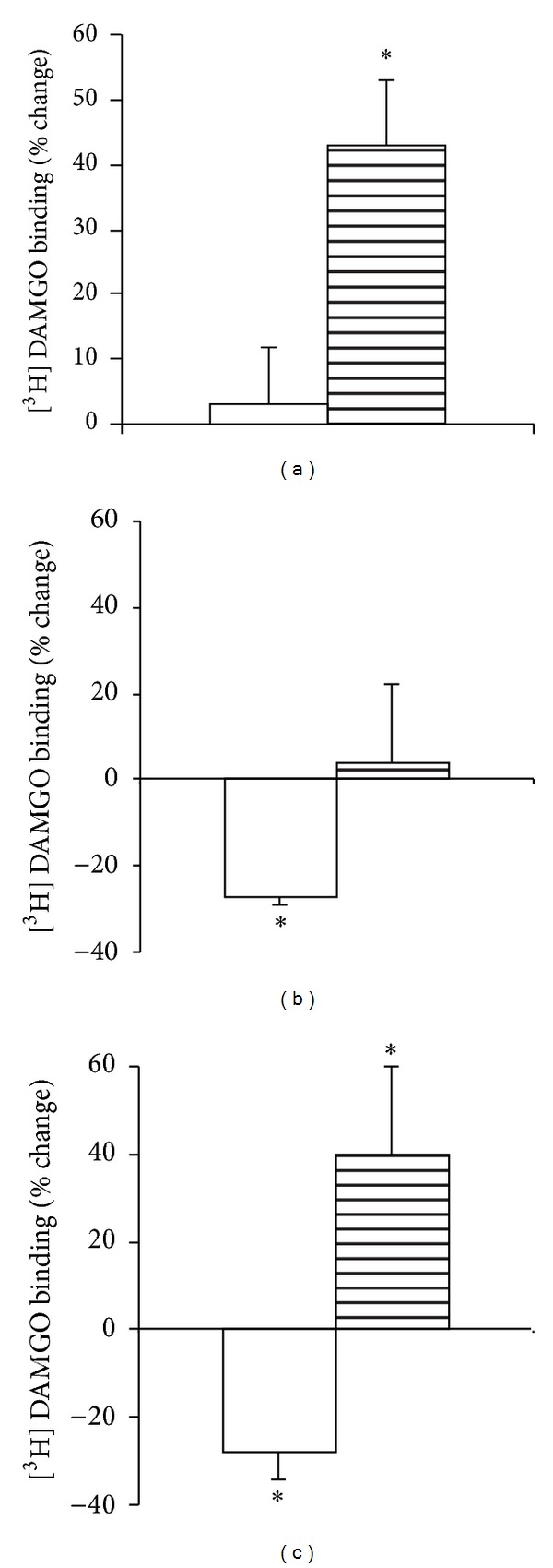
Changes in receptor density (B max) following chronic exposure of ACHC-EM2 (a) DAMGO (b), and DAMCK (c). SPM (white columns) and MI (striped columns) fractions were prepared from whole brain. The membrane suspensions (0.3 mg protein) were incubated with 1 nM [3H]DAMGO for 60 min at 25°C in the absence (total binding) or in the presence of 10−10–10−5 M of unlabeled DAMGO. Results are expressed as % change of protein in each fraction. Mean ± S.E.M.; n = 3–6; significance was determined by t-test; *P < 0.05 compared to control.