
Figure 1.
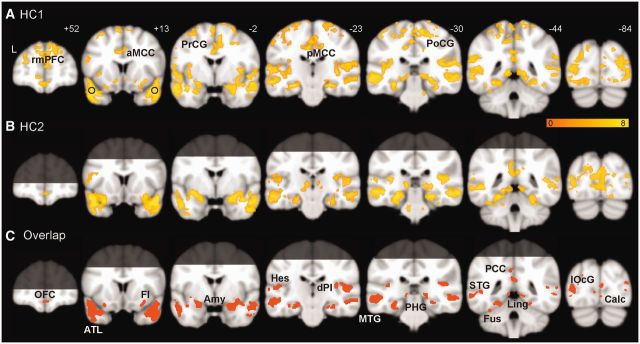
Intrinsic ATL connectivity in the healthy brain. Group-level ATL-seeded intrinsic connectivity network maps are shown for HC1 (A), HC2 (B), and their overlap (C), generated with a joint height-extent threshold (P < 0.0001 for peak height and FWE-corrected P < 0.05 for spatial extent). Brain regions beyond the coverage of the optimized EPI protocol are shaded in B and C, and thus only labelled in A. Black circles in A signify the location of the ATL seeds. Amy = amygdala; aMCC = anterior midcingulate cortex; Calc = calcarine; dPI = dorsal posterior insula; FI = frontoinsula; Fus = fusiform; Hes = Heschl’s; Ling = lingual gyrus; lOcG = lateral occipital gyrus; MTG = middle temporal gyrus; OFC = orbitofrontal cortex; PCC = posterior cingulate cortex; pMCC = posterior midcingulate cortex; PCu = precuneus; PHG = parahippocampus; PoCG = postcentral gyrus; PrCG = precentral gyrus; rmPFC = rostral medial prefrontal cortex; STG = superior temporal gyrus.