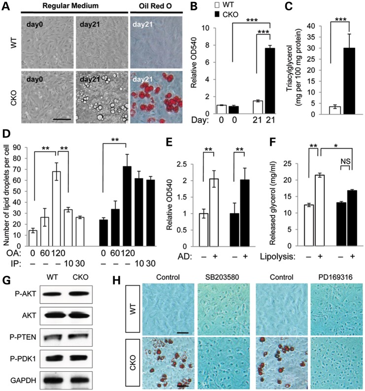
Figure 1.
Spontaneous accumulation of lipid droplets in Tgfbr2 mutant cells resulting from a defect in lipolysis. (A) Primary MEPM cells from Tgfbr2fl/fl (WT) and Tgfbr2fl/fl;Wnt1-Cre (CKO) mice cultured in regular medium for 0 or 21 days. Lipid droplets were stained with Oil Red O. Bar, 50 μm. (B) Quantitative analysis of lipid accumulation by Oil Red O staining in WT (white bar) and CKO (black bar) MEPM cells. Data are OD490 measurements. ***P<0.001. (C) Triacylglycerol level in WT and CKO MEPM cells cultured for 21 days. Data are expressed as mg per 100 mg protein. ***P < 0.001. (D) Pulse-chase analysis of lipid droplet formation using chemical inducers, oleic acid (OA) for lipogenesis (after 0, 60 and 120 min) and isoproterenol (IP) for lipolysis (after 10 and 30 min), in MEPM cells of WT and CKO mice. **P < 0.01. (E) Quantitative analysis of adipogenesis by Oil Red O staining in WT and CKO MEPM cells after 1 week of culture in adipogenic induction (+) or regular medium (−). Data are relative OD490 measurements. **P < 0.01. (F) Quantitative analysis of glycerol released into the medium by isoproterenol stimulation (+) or mock treatment (−) in MEPM-derived adipocytes of WT and CKO mice. *P < 0.05; **P < 0.01; NS, not significant. (G) Immunoblotting analysis of indicated molecules related to AKT signaling in MEPM cells from WT and CKO mice. (H) Oil Red O staining of MEPM cells from WT and CKO mice treated without (control) or with p38 MAPK inhibitor SB203580 or PD169316 for 3 weeks. Bars, 20 μm.

