
Figure 3.
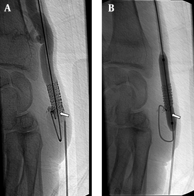
A 53-year-old man with radiocephalic fistula in the left forearm. He had a previous stent placement in this area. A, AV fistulogram shows the thrombus in the stent that occurred in the third month follow-up (arrow). B, The balloon dilatation (6 mm) is used for the stent expandation and a new 7 mm sized second stent is placed at the same area. The control angiogram images show patency of the vascular access (arrow).