
Figure 4.
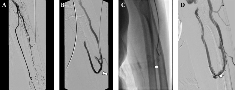
A 75-year-old man with radiocephalic access in the left forearm in place at the third day after PTA; A, Fistulogram shows occlusion of the distal radial artery. B, The metallic stent 6×40 mm deployed at the radial artery and the cephalic vein; the fistulogram shows the blood supply at this access (arrow). C, The follow-up venogram obtained after 8 months with the retrograde catheterization shows laceration of the stent (arrow). D, Angiogram shows patency of the stent and the access (arrow), an additional interventional procedure is not required.