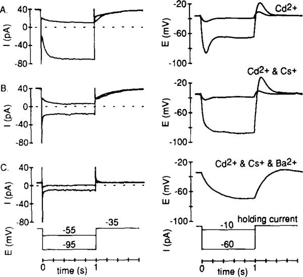
Figure 6. Effects of IKx and Ih on the Voltage Response to Current Injection.
Current components were identified in the voltage-clamp mode, and voltage responses to square pulses of injected current were assessed in the same cell. In each condition, cells were made to “rest” at about –38 mV by injection of a constant holding current (Axopatch 1-C).
(A) To reveal Ih, recordings were made in the absence of Cs+ but in the presence of Cd2+. With both Ih (dominant at –95 mV) and IKx (dominant at –55 mV) present, a time-dependent decrease in both small and large voltage signals occurred.
(B) Recordings in both (B) and (C) were made from cells bathed in Ringer solution containing 5 mM Cs+ and 0.1 mM Cd2+. Bath application of 5 mM Cs+ blocked Ih, but left IKx. The small voltage signal was similar to the control but the large signal became sustained.
(C) Bath-applied 5 mM Ba2+ inhibited IKx in this voltage range (see Figure 5). No time- or voltage-dependent currents (other than capacity currents) were evident, and the response to the small hyperpolarizing current injection was much larger and did not decrease with time (the larger hyperpolarizing current step was not applied).

