Fig. 1.
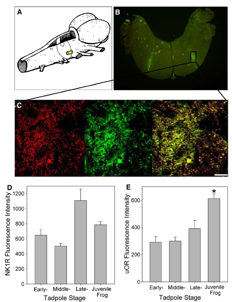
NK1R and µOR colocalization in the juvenile bullfrog brainstem. a Drawing illustrates the approximate location and size of NK1R and µOR colocalization in the juvenile bullfrog brainstem. b NK1R and µOR colocalize on the ventral surface of the brainstem near the facial nucleus in juvenile bullfrog brainstems (n = 8). c Magnified view of NK1R (red) and µOR (green) colocalization (yellow) in the region of interest. The bar indicates 10 µm. d NK1R fluorescence intensity has no developmental trend (n = 6–8). e µOR fluorescence intensity increased significantly from late-stage tadpole to juvenile bullfrog (n = 6–8). Data are mean ± SEM. Asterisk indicates a significant difference from all other groups (P < 0.05)

