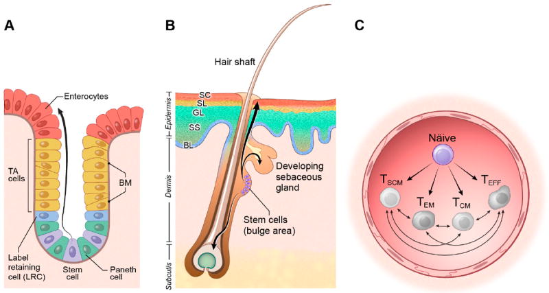
Figure 1. The linage relationship of T cell subsets is complicated by the lack of anatomical clues.

A) The intestinal crypt-villus unit. Intestinal stem cells reside at the base of the crypt between Paneth cells. As cells proliferate and differentiate into transient amplifying (TA) progenitor cells and mature enterocytes, they move upwards to cover the villus. B) The skin. Epidermal stem cells are located in the bulge region of the hair follicle, the base of the sebaceous gland, and the basal layer of the interfollicular epidermis. As cells proliferate and differentiate into keratinocytes, they move upward to form the stratum spinosus (SS), the granular layer (GL), the stratum lucidum (SL) and the stratum corneum (SC). C) T cells. Following antigen-stimulation, naïve T cells differentiate generating the full diversity T cell subsets. The existence of cells at different developmental stages, all of which are moving within the same anatomical space, does not offer an easy static snapshot that provides clues about their lineage relationships which are still the subject of controversy in the field.
