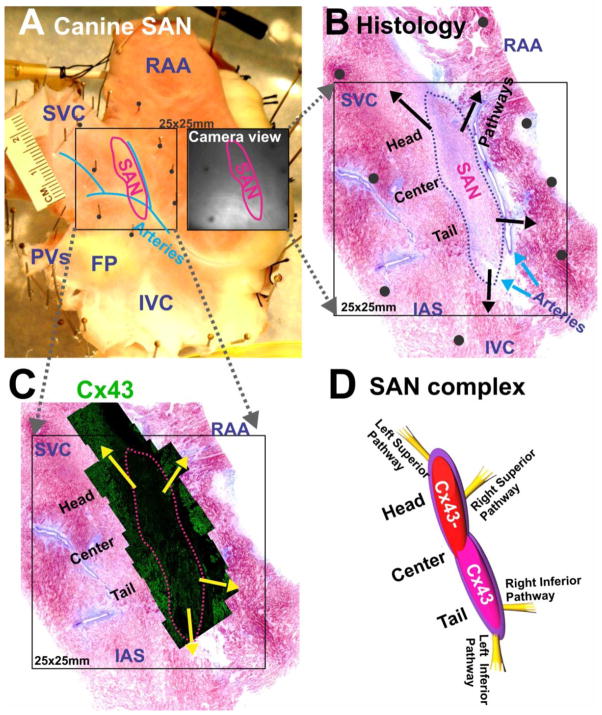
Figure 1. Experimental preparation and histology of the canine sino-atrial node (SAN).
(A) An epicardial view of a canine SAN preparation. The SAN is demarcated by a red line. The black square shows the mapped area containing the SAN. The adjacent black-and-white image shows the same area seen from a camera. (B) A parallel histology section of SAN close to the epicardial surface. Arrows indicate locations of SAN conduction pathways. Black dots indicate locations of pins in panel A. (C) Immunolabeling of connexin 43 (Cx43) of SAN confirms the boundary of functionally defined SAN. (D) A graphic representation showing the SAN complex and the superior and inferior SACPs.
SVC: superior vena cava; IVC: inferior vena cava; RAA: right atrial appendage; PVs: pulmonary veins; FP: fat pad; IAS: interatrial septum.

