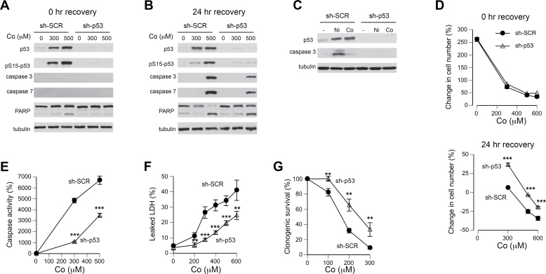
FIG. 4.
Role of p53 in cytotoxicity of Co(II) in H460 cells. A, Western blots for samples collected at the end of 24-h treatments with Co(II) (sh-SCR—cells expressing nonspecific shRNA and sh-p53—cells with p53-targeting shRNA). B, Western blots for samples collected at 24h posttreatment. C, Western blots for p53 and cleaved (active) caspase-3 in cells treated with 250μM Ni(II) or Co(II) for 48h. D, Effect of p53 depletion on growth inhibition by Co(II) at the end of 24-h treatments (top panel) and following 24-h recovery in metal-free media (bottom panel). Means ± SD for 4 independent determinations. ***p < 0.001 relative to the same concentration in sh-SCR samples. Error bars were smaller than data symbols in most cases. E, Caspase 3/7 activity in H460 cells following 24-h recovery in Co-free media. Means ± SD for 4 independent determinations. ***p < 0.001 relative to the same concentration in sh-SCR samples. F, LDH leakage from H460 cells during 24h posttreatment incubation. Means ± SD for 4 independent determinations. **p < 0.01 and ***p < 0.001 relative to the same concentration in sh-SCR samples. G, Clonogenic survival of H460 cells treated with Co(II) for 24h. Means ± SD for 3 independent determinations. **p < 0.01 relative to the same concentration in sh-SCR samples.

