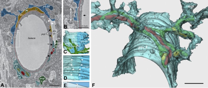
Figure 3.
The perivascular astroglial sheath provides a complete covering of brain microvessels. A: electron microscopic image of capillary with four endfoot profiles (pve I–IV). The hatched area (enlarged in B) shows endfoot-endfoot overlap. The double arrow in B represents the projection of the overlap, while the black arrow indicates the angle of view in C. C: 3D reconstruction. Endfoot IV is removed to view the remaining endfeet from the inside of the vessel. In the hatched frame, enlarged in D, endfoot I (pve I) is made semitransparent to visualize the underlying endfoot III (pve III). The space between the two rows of arrowheads represents the extent of overlap. E: definition of the intercellular cleft length (IC) and the length of its projection (P). F: a semitransparent 3D reconstruction of a perivascular endfoot (pve I) seen from an abluminal perspective. The footplate is complete and covers about half the circumference of the vessel wall. Elongated mitochondria (green, red, and yellow) enter the endfoot tangentially through the left process (asp) and leave through four processes on the right hand side. Scale bar = 1 μm unless otherwise indicated. [Modified from Mathiisen et al. (101), copyright 2010. This material is reproduced with permission of John Wiley & Sons, Inc.]

