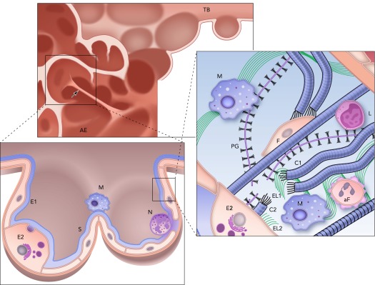
FIGURE 2.

Structure and complexity of the parenchyma at three length scales in the emphysematous lung
Top: the remnants of a terminal bronchiole (TB) leading to airspace enlargement (AE). Note the single straight septal wall under larger than average mechanical stress (arrow). Bottom left: zoomed in image of an enlarged airspace region that used to be two alveoli lined with type I (E1) and type II (E2) epithelial cells covered by a thin liquid layer and surfactant (S). Inflammatory cells, including a macrophage (M) and a neutrophil (N), are also shown. Right: a schematic representation of the extracellular matrix of the septal wall with various components including amorphous elastin (El) and collagen (C), and reduced amount of proteoglycans (PG) and fibroblast (F). Compared with the normal ECM in FIGURE 1, here two macrophages (M) and an apoptotic fibroblast (aF) are also seen. Additionally, collagen C is straight because it is under tension, whereas the wavy fragments of collagen C1 and C2 show a rupture site, where elastin (El1 and El2) as well as PG fragments are found. Note also that a macrophage is recruited near this rupture site by the fragments. L denotes a lymphocyte.
