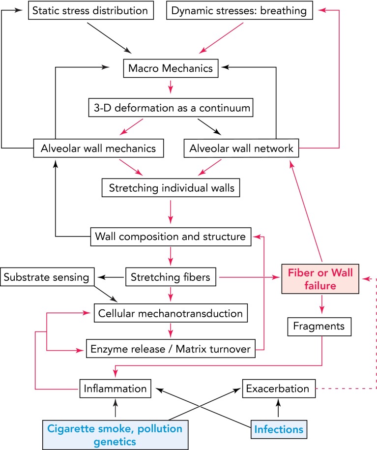
FIGURE 5.
Schematic diagram showing the complexity of multiscale signaling and mechanics with embedded feedback mechanisms influencing ECM composition and lung function in emphysema
The arrows represent known or possible links. Notice the feedback from alveolar wall and network mechanics to static stress distribution and dynamic stresses, which decrease transpulmonary pressure and alter breathing pattern. The blue at the bottom represents internal or external triggers. The red pathways show a possible mechanism of self-sustained progression. Fiber and wall failure (red) play a key role here since they directly generate airspace enlargement, feed back to breathing pattern, and, through exposure of fragments, maintain inflammation and drive signaling. The red dashed line represents the added effect of exacerbations on the steady progression.

