
Figure 2. The Western blotting results of cell lines were consistent with the Immunohistochemical analysis of gastric cancer tissues.
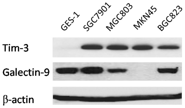
Note that both Tim-3 and Gal-9 were detected in gastric cancer cell lines MGC803,SGC7901, and BGC823, absence of Gal-9 activation in MKN45 cells, and negative of Tim-3 immunostaining in GES-1 were also confirmed.