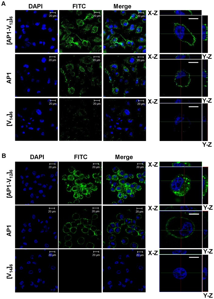
Figure 4. Analysis of cellular localization of [AP1-V12]6 polymer.
Confocal laser scanning microscopic images of H226 cancer cells treated with 10 µM of [AP1-V12]6, AP1, or [V14]6 at (A) 4°C and (B) 37°C. Representative confocal images of three experiments (scale bar 20 µm). Right panels: Examination of [AP1-V12]6, AP1 and [V14]6 cellular location by Z-section scanning of confocal microscopic images. Representative confocal images of three experiments (scale bar 10 µm).

