
Figure 2. His-Tag purification of phosphorylated ADAP (ADAP-P) and non-phosphorylated ADAP (ADAP-OH).
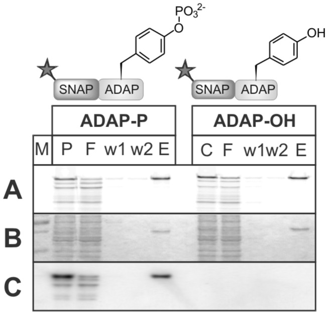
(A) Detection of fluorescent ADAP in SDS gel (excitation 633 nm), (B) Coomassie stain, (C) Western Blot with anti phosphotyrosine antibody. P: cell-free synthesis of ADAP after in vitro phosphorylation, C: non-phosphorylated ADAP synthesis, F: flow-through, w1 and w2: wash fractions, E: purified ADAP.