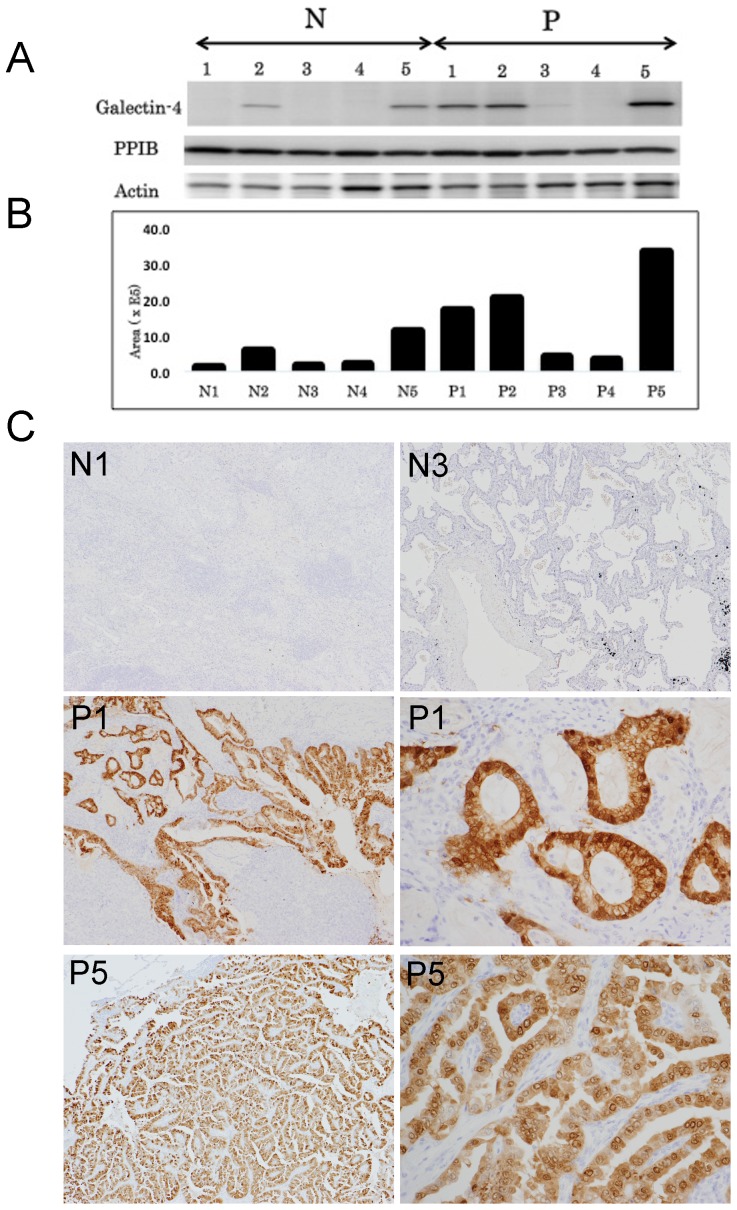
Figure 1. Validation of the differential expression of galectin-4.
A. A representative result of western blotting for galectin-4 expression and PPIB, together with β-actin as internal control. Three of 5 lung adenocarcinomas with LN metastasis expressed galectin-4 at higher levels than those without LN metastasis. One lung adenocarcinoma with LN metastasis (P3) showed a slightly increased expression level of galectin-4. PPIB expression was observed almost equally in all samples examined. B. Histogram showing the relative expression levels of galectin-4. C. Immunohistochemical expression patterns of galectin-4 in normal lung tissue and lung adenocarcinoma tissues. Lung adenocarcinomas without lymph node metastasis (N1 and N3) showed that both carcinomas and normal lung tissue did not express galectin-4, whereas lung adenocarcinomas with lymph node metastasis (P1 and P5) showed that galectin-4 was strongly expressed in carcinoma cells (magnification ×20). The high-power view of lung adenocarcinomas with lymph node metastasis showed nuclear, cytoplasmic, and membranous expression of galectin-4 (magnification ×100).

