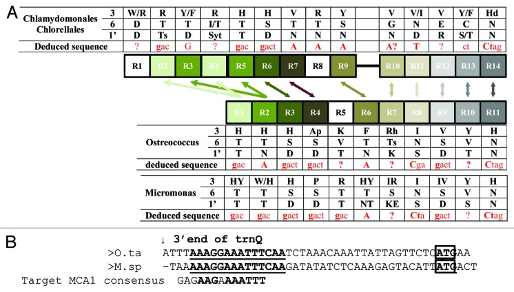
Figure 5. (A) Schematic comparison of the distribution of the PPR repeats in the MCA1 protein of Chlorophyceae vs. Trebouxiophyceae. The phylogenetically related repeats are connected by arrows. Key residues critical for the nucleotide recognition are indicated, as well as the putative target sequence (in red) as deduced from the ?PPR code.? (B) Alignment of the petA 5?UTRs from O. tauri and M. pusilla RCC299. The arrow points to the 3? end of the trnQ, located immediately upstream of petA and the petA initiation codon is boxed. A region conserved between the two organisms is written in bold and underlined. The MCA1 binding site in Chlamydomonas is shown below for comparison.

An official website of the United States government
Here's how you know
Official websites use .gov
A
.gov website belongs to an official
government organization in the United States.
Secure .gov websites use HTTPS
A lock (
) or https:// means you've safely
connected to the .gov website. Share sensitive
information only on official, secure websites.
