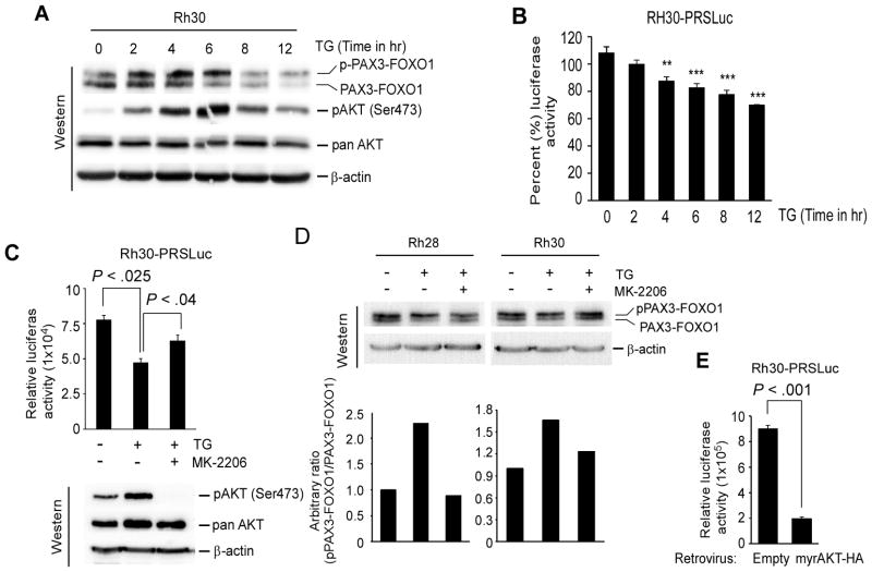
Figure 2.
Thapsigargin mediates AKT activation can increase PAX3-FOXO1 phosphorylation and inhibit its transcriptional activity in ARMS cells. (A) Immunoblot of extracts from Rh30 cells incubated with 8.0 nM TG for indicated time points, probed for PAX3-FOXO1, activated pAKT (Ser473), total AKT, and β-actin for loading control. (B) Luciferase activity in Rh30-PRSLuc reporter cells treated with 8.0 nM TG for indicated time points. Values were normalized to protein. Error bars, ± SEM, (n=3). P-value ** = 0.001 to 0.01, *** = 0.0001 to 0.001. (C) Luciferase activity in Rh30-PRSLuc cells treated with vehicle control or 8.0 nM TG along with or without AKT inhibitor MK-2206 (1.0 μM) for 4 hours. Values were expressed after normalization with protein. Error bars, ± SEM, (n=3). (top). Immunoblot of cell extracts probed for activated pAKT (Ser473), total AKT, and β-actin for loading control (bottom). (D) Immunoblot of extracts from indicated cells treated with TG along with or without MK-2206 as described in (C), probed with FOXO1 and β-actin antibodies (top). Relative level of phosphorylated PAX3-FOXO1 (upper) is represented as a bar graph (bottom). (E) Luciferase activity in Rh30-PRSLuc cells transduced with retrovirus expressing a constitutively active AKT (myrAKT-HA) or control retrovirus (empty) grown for 2 days. Values were expressed after protein normalization. Error bars, ± SEM, (n=3).

