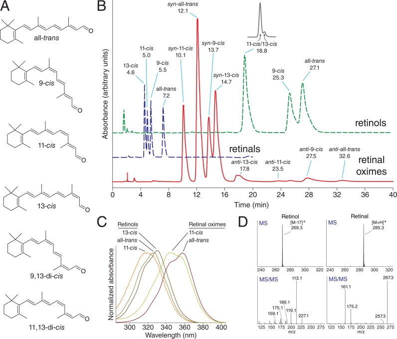
Figure 6.
HPLC-based separation and detection of retinoids. (A) The main classes of retinoid isomers commonly found in experimental samples that can be distinguished by analytical methods. (B) Elution profiles of retinol, retinal, and retinal oxime isomers from a normal phase HPLC column with an isocratic flow of 10% ethyl acetate/hexane. Primary analytical methods of retinoid identification and quantification include UV/vis spectroscopy and mass spectrometry. (C) UV/vis absorbance spectra of selected retinoids reveal characteristic differences in absorbance maxima and overall shape of the spectra that are used to classify the chemical and geometric form of retinoids. (D) Electrospray ionization of retinol and retinyl esters triggers water or carboxylate dissociation, resulting in the predominant parent ion of m/z = 269 [M – 17]+, whereas retinal exhibits the expected molecular ion of m/z = 285 [M + H]+. Characteristic MS/MS fragmentation patterns of the parent ions are shown in the bottom panels. The typical m/z = 161 fragment of retinal in MS/MS spectra is indicative of ionone ring loss from the parent ion.

