Figure 1.
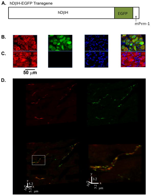
Generation of hDβH-EGFP transgenic mice. A, Schematic diagram of the hDβH-EGFP transgene. mPrm-1 denotes murine protamine-1. B, Portion of a midsection through a stellate ganglion from an adult hDβH-EGFP transgenic mouse (line 4) stained for expression of tyrosine hydroxylase (TH; red) and EGFP (green), and counterstained with nuclear Hoechst (blue). The three-channel merged image is shown in the right panel. TH-expressing cells exhibit both cytoplasmic and nuclear anti-EGFP immunofluorescence. C, Section through a control stellate ganglion stained as in the transgenic ganglion, showing TH-expressing and non-expressing cells. D, Volume renderings of a 30-slice confocal z-stack obtained from a 10-μm-thick section from the left ventricular free wall of an adult hDβH-EGFP heart (line 4) dually stained with an antiTH and an anti-GFP antibody, followed by exposure to Alexa546- (red) and Alexa633- (green) conjugated secondary antibodies. The yellow color in the merged image indicates co-localization of TH and EGFP. The right panel is a magnified view of the boxed region in the merged image.

