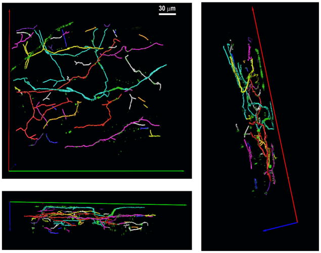
Figure 6.
Tiled arrangement of sympathetic subarbors in the left ventricular subepicardium. Volume-rendered trajectories were obtained for each invidually identified subarbor using the 3-D tracing algorithm and assigned contrasting colors. The 3-D color map is shown from three different view angels.

