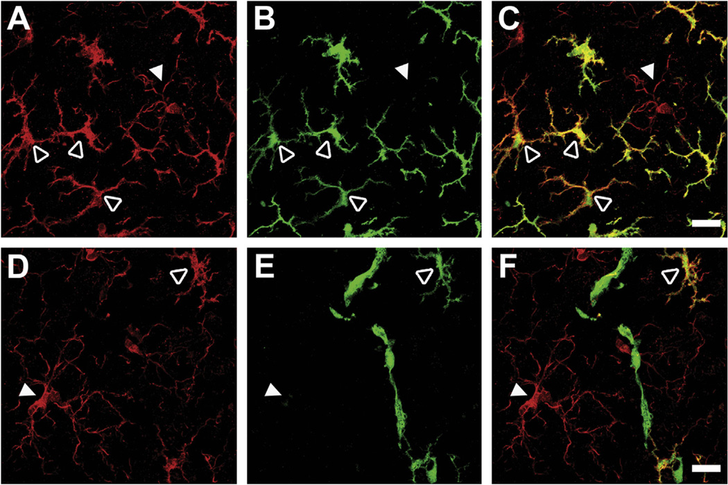
Fig. 6.
Morphology of GFP+ cells recruited to the CNS. Confocal microscopy of CNS sections from recipient mice confirmed that GFP+ cells expressed microglial marker Iba1 and had a stellate appearance (A–F, open arrowheads) compared with resident microglia which displayed a ramified (“resting”) phenotype (A–F, closed arrowheads). Representative images showing microglia in red (Iba1; A and D), GFP in green (B and E), and overlay in yellow-orange (C and F). Co-localization of GFP and Iba1 staining was used to distinguish recruited cells (Iba1+GFP+) from resident microglia (Iba1+GFP−) (upper panels). The few GFP+ cells that were negative for Iba1 were associated with blood vessels with an elongated morphology similar to perivascular macrophages (lower panels). Scale bars = 20 µm. (For interpretation of the references to colour in this figure legend, the reader is referred to the web version of this article.)

