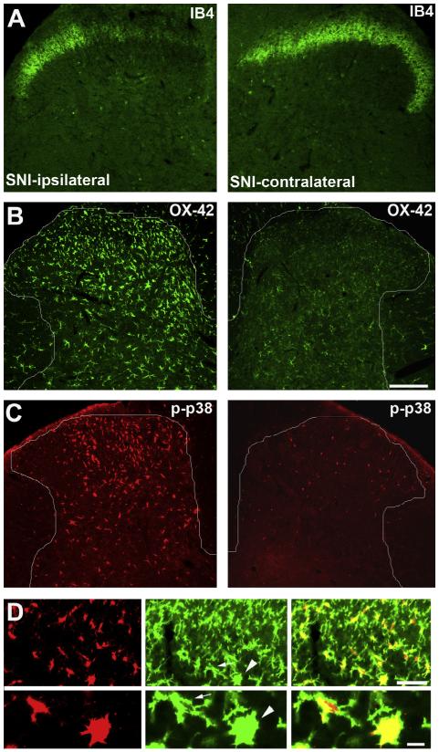
Fig. 2.
Activation of microglia in the spinal cord dorsal horn 3 days after spared nerve injury (SNI) in rats. (A) IB4 staining in the spinal cord dorsal horn ipsilateral and contralateral to the injury side. Note a loss of IB4 staining in the dorsal horn region innervated by the injured nerve branches. (B and C) CD11b (OX-42) and phosphorylated p38 (p-p38) immunostaining in the dorsal horn ipsilateral and contralateral to the injury side. Note overlapping expression patterns of OX-42 and p-p38 in the injury side. (D) Double staining of p-p38 (red) and OX-42 (green) in the ipsilateral dorsal horn. Lower panel presents high-magnification images of 2 microglial cells (indicated by arrow and arrowhead) from the upper panel. Note that p-p38 is completely co-localized with OX-42. Scale, 100 lm. Images are modified from Wen et al. [276], with permission.

