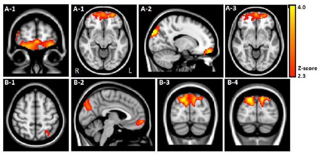
Figure 1.
VBM results contrasting PD < Control indicated GM volume loss in PD subjects in (A) Frontal cortex: bilateral frontal pole (A-1; BA 10, 11) encompassing the orbitofrontal cortex (A-2) and bilateral medial frontal gyrus (A-3; BA 10, 32); (B) Parieto-occipital cortex shown in axial, sagittal, and coronal planes: 1) left superior parietal cortex (BA 7), 2) bilateral parieto-occipital junction (BA 7, 18, 19), 3) bilateral lateral occipital cortex (BA 19), and 4) bilateral superior occipital cortex (BA 19).

