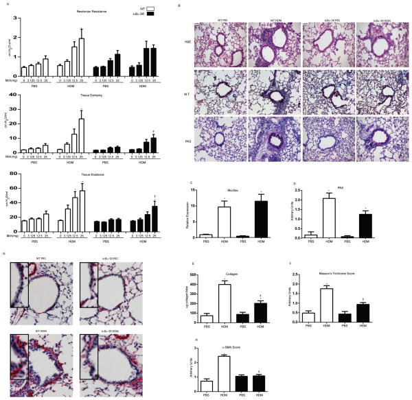
Figure 5. Assessment of HDM-induced alterations in respiratory mechanics and airway remodeling in WT or CC10-NF-κBSR mice following 15 challenges with HDM.
(A) Evaluation of airway hyperresponsiveness via forced oscillation mechanics in response to 3.125, 12.5, and 25 mg methacholine. Shown are the parameters Newtonian resistance (Rn), Tissue damping (G) and Elastance (H) assessed via forced oscillation (B) Histopathological evaluation of tissue inflammation (H&E), mucus metaplasia (PAS) and peri-bronchiolar collagen deposition (M.T.). scale bar=50μm. (C) Analysis of Muc5ac mRNA expression in homogenized lung tissue. Data were normalized to cyclophilin and are presented as relative expression. (D) Quantification of mucus metaplasia. Bronchioles of similar size (n=3) were analyzed/mouse by two blinded scorers, and average scores presented as average units. (E) Assessment of collagen from the upper right lobe. (F) Quantification of peri-bronchiolar collagen deposition. Bronchioles of similar size (n=3) were analyzed/mouse by two blinded scorers, and average scores presented as average units. (G) Assessment of α-SMA immunohistochemistry, red=α-SMA, scale bar=1μm. (F) Quantification of α-SMA immunoreactivity. Bronchioles of similar size (n=3/mouse) were analyzed by two independent blinded scorers, and average scores presented as average units. Data represent 8 mice/group/time point. * p< 0.05 (ANOVA) compared to the PBS group at the same time point. † p<0.05 (ANOVA, Kruskal Wallis) compared to WT mice exposed to HDM.

