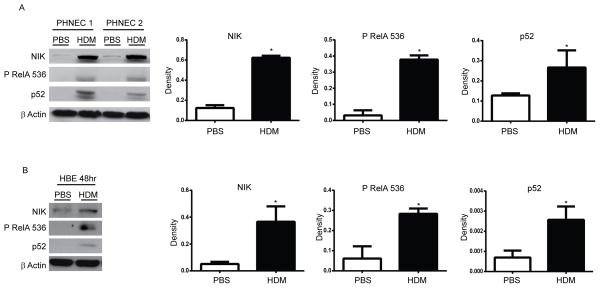
Figure 6. Assessment of activation of classical and alternative NF-κB pathways in human lung epithelial cells exposed to HDM.
(A) Primary human nasal epithelial (PHNE) cells or (B) Human bronchial epithelial (HBE) cells were exposed to PBS or 25 μg HDM once a day for consecutive days and harvested thereafter at either 72 h (PHNE) or 48 h (HBE). Cells were lysed for evaluation of NIK, p52, phospho RelA 536, by Western blot analyses. β-Actin (loading control). Right panels: Densitometric evaluation of Western blots shown in (A) PHNE (n=3 patients) or (B) HBE (n=3 experimental repeats). Results are expressed as arbitrary density and were normalized to corresponding β-Actin bands.

